


|
|
What are Obstetric Ultrasound
Scans?
Obstetric Ultrasound is the use of ultrasound scans in pregnancy. Since
its introduction in the late 1950’s ultrasonography has become a very
useful diagnostic tool in Obstetrics. Currently used equipments are known
as real-time scanners, with which a continous picture of the moving fetus
can be depicted on a monitor screen. Very high frequency sound waves of
between 3.5 to 7.0 megahertz (i.e. 3.5 to 7 million cycles per second)
are generally used for this purpose. They are emitted from a transducer
which is placed in contact with the maternal abdomen, and is moved to
"look at" (likened to a light shined from a torch) any particular content
of the uterus. Repetitive arrays of ultrasound beams scan the fetus in
thin slices and are reflected back onto the same transducer. The information
obtained from different reflections are recomposed back into a picture
on the monitor screen (a sonogram, or ultrasonogram). Movements such as
fetal heart beat and malformations in the feus can be assessed and measurements
can be made accurately on the images displayed on the screen. Such measurements
form the cornerstone in the assessment of gestational age, size and growth
in the fetus.
A short history of the development
of ultrasound in pregnancy can be found in the History
pages.
Why and
when is Ultrasound used in Pregnancy?
Ultrasound scan is currently considered to be a safe, non-invasive, accurate
and cost-effective investigation in the fetus. It has progressively become
an indispensible obstetric tool and plays an important role in the care
of every pregnant woman. The main use of ultrasonography are in the following
areas:
1. Diagnosis and assessment of early pregnancy. The gestational sac can
be visualized as early as four and a half weeks of gestation and the yolk
sac at about five weeks.
2. Threatened miscarriage. The viability of the fetus can be documented
in the presence of vaginal bleeding in early pregnancy. Fetal heart motion
is usually clearly depictable by 7 weeks. If this is observed, the probability
of a continued pregnancy is greater than 97 percent. Missed abortion and
blighted ovum will usually give typical pictures of a deformed gestational
sac and absence of fetal poles or heart beat. Ultrasonography is also
indispensible in the early diagnosis of ectopic pregnancies and molar
pregnancies.
3. Determination of gestational age and assessment of fetal size. Fetal
body measurements reflect the gestational age of the fetus. This is particularly
true in early gestation. In patients with uncertain last menstrual periods,
such measurements must be made as early as possible in pregnancy to arrive
at a correct dating for the patient. In the latter part of pregnancy measuring
body parameters will allow assessment of the size and growth of the fetus
and will greatly assist in the diagnosis and management of intrauterine
growth retardation (IUGR).
The following measurements are usually made:
a) The Crown-rump length (CRL) This measurement can be made between 7
to 13 weeks and gives very accurate estimation of the gestational age.
Dating with the CRL can be within 3-4 days of the last menstrual period.
(Table)
b) The Biparietal diameter (BPD) The diameter between the 2 sides of the
head. This is measured after 13 weeks. It increases from about 2.4 cm
at 13 weeks to about 9.5 cm at term. Different babies of the same weight
can have different head size, therefore dating in the later part of pregnancy
is generally considered unreliable. (Chart and further comments)
c) The Femur length (FL) Measures the longest bone in the body and reflects
the longitudinal growth of the fetus. Its usefulness is similar to the
BPD. It increases from about 1.5 cm at 14 weeks to about 7.8 cm at term.
(Chart and further comments)
d) The Abdominal circumference (AC) The single most important measurement
to make in late pregnancy. It reflects more of fetal size and weight rather
than age. Serial measurements are useful in monitoring growth of the fetus.
(Chart and further comments) Other important measurements are discussed
here. The weight of the fetus at any gestation can also be estimated with
great accuracy using polynomial equations containing the BPD, FL, and
AC. Lookup charts are readily available. For example, a BPD of 9.0 cm
and an AC of 30.0 cm will give a weight estimate of 2.85 kg. (comments)
4. Placental localization. Ultrasonography has become indispensible in
the diagnosis or exclusion of placenta previa, and other placental abnormalities
as in diabetes, fetal hydrops, Rh isoimmunization and severe intrauterine
growth retardation .
5. Multiple pregnancies. In this situation, ultrasonography is invaluable
in determining the number of fetuses, the chorionicity, fetal presentations,
evidence of growth retardation and fetal anomaly, the presence of placenta
previa, and any suggestion of twin-to-twin transfusion.
6. Hydramnios and Oligohydramnios. Excessive or decreased amount of liquor
(amniotic fluid) can be clearly depicted by ultrasound. In both these
situations, careful ultrasound examination should be made to exclude intraulterine
growth retardation and congenital malformation in the fetus such as intestinal
atresia, hydrops fetalis or renal dysplasia. See FAQ and comments.
7. Fetal malformation. Many structural abnormalities in the fetus can
be reliably diagnosed by an ultrasound scan, and these can usually be
made before 20 weeks. Common examples include hydrocephalus, anencephaly,
myelomeningocoele, achondroplasia and other dwarfism, spina bifida, exomphalos,
duodenal atresia and fetal hydrops. With more recent equipments, conditions
such as cleft lips/palate, congenital cardiac abnormalities and Down syndrome
are more readily recognised. Markers for chromosomal abnormalities such
as the fetal nuchal translucency (the area at the back of the neck) have
also been defined to enable detection of these abnormal fetuses. Ultrasound
can also assist in other diagnostic procedures in prenatal diagnosis such
as amniocentesis, chorionic villus sampling, percutaneous umbilical blood
sampling and in fetal therapy.
8. Other areas. Ultrasonography is of great value in other obstetric conditions
such as:
a) confirmation of intrauterine death.
b) confirmation of fetal presentation in uncertain cases.
c) evaluating fetal movements, tone and breathing in the Biophysical Profile.
d) diagnosis of uterine and pelvic abnormalities during pregnancy e.g.
fibromyomata and ovarian cyst.
The Schedule

There is no hard and fast rule as to the number of scans a woman should
have during her pregnancy. A scan is ordered when an abnormality is suspected
on clinical grounds. Otherwise a scan is generally booked at about 7 weeks
to confirm pregnancy, exclude ectopic or molar pregnancies, confirm cardiac
pulsation and measure the crown-rump length for dating. A second scan
is performed at 18 to 20 weeks to look for congenital malformations, exclude
multiple pregnancies and to verify dates and growth. Placental position
is also determined. A third scan may sometimes be done at around 34 weeks
to evaluate fetal size and assess fetal growth. Placental position is
verified. Many centers are now doing a scan at around 13-14 weeks to measure
the nuchal skin fold thickness for the purpose of evaluating the risk
for Down Syndrome. The total number of scans will vary depending on whether
a previous scan has detected certain abnormalities that require follow-up
assessment. What is often referred to as a Level II scan merely indicates
a "targeted" examination where it is done when an indication is present
or when an abnormality is suspected in a previous examination. In fact
professional bodies such as the American Institute of Ultrasound in Medicine
does not endorse or encourage the use of these terms. A more "thorough"
examination is usually done at an a perinatal center or specialised clinic
where more expertise and better equipments may be present. One should
not dwell too much on the definitions or guidelines for a level II ultrasound
scan. The sonologist should always try very hard to look for and assess
any abnormality that may be present in the fetus. It is not very meaningful
to be talking about level III or even level IV scans. Whether a pregnancy
must be scanned on a 'routine' basis at 18 to 20 weeks is still a matter
of some controversy.
Transvaginal
Scan
With specially designed probes, ultrasound scanning can be done with the
probe placed in the vagina of the patient. This method usually provides
better images (and therefore more information) in patients who are not
pregnant or are in the early stages of pregnancy. Fetal cardiac pulsation
can be observed as early as 6 weeks of gestation. Vaginal scans are becoming
indispensible in the early diagnosis of ectopic pregnancies. An increasing
number of fetal abnormalities are now being diagnosed in the first trimester
using the vaginal scan. Transvaginal scans on the other hand are also
useful in the second trimester in the diagnosis of congenital anomalies.
Read one of my presentations at OBGYN.net-Ultrasound.
Doppler
Ultrasound
The doppler shift principle has been used for a long time in fetal heart
rate detectors. Further developments in doppler ultrasound technology
in recent years have enabled a great expansion in it's application in
Obstetrics, this time in the area of assessing and monitoring the well-being
of the fetus. Blood flow characteristics in the fetal blood vessels can
be assessed with Doppler 'flow velocity waveforms'. Diminished flow, particularly
in the diastolic phase of a pulse cycle is associated with compromise
in the fetus. Various ratios of the systolic to diastolic flow are used
as a measure of this compromise. The blood vessels commonly interrogated
include the umbilical artery, the aorta, the middle cerebral arteries
and the uterine arcuate arteries. The use of color flow mapping can clearly
depict the flow of blood in fetal blood vessels in a realtime scan, the
direction of the flow being represented by different colors. 'Color' doppler
is particularly indispensible in the diagnosis and assessment of congenital
heart abnormalities. Another recent development is the Power Doppler (Doppler
angiography). It uses amplitude information from doppler signals rather
than flow velocity information to visualize slow flow in smaller blood
vessels. A color perfusion-like display of a particular organ such as
the placenta overlapping on the 2-D image can be very nicely depicted.
Doppler examinations can be performed abdominally and via the transvaginal
route. The power emitted by a doppler device is generally greater than
that used in a conventional 2-D scan.
Color
imaging
This is a recent addition to plain 2-D realtime scanning. Also known as
"chroma" scans, user-selectable color hues are assigned to the shades
of grey for better visualization of subtle tissue details. This clever
enhancement is aimed at better interpretation of the scans. 'Color scans'
do not imply that various parts of the same picture are depicted in different
colors like what we see in a color photograph.
3-D Ultrasound
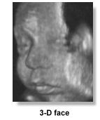
3 dimensional ultrasound is quickly moving out of the research and development
stages and is very much in the News. Faster and more advanced commercial
models are coming into the market. The scans requires special probes and
software to accumulate and render the images, and the rendering time has
been reduced from minutes to seconds. A good 3D image is often quite impressive
and further 2D scans may be extracted from 3D blocks of scanned information.
Volumetric measurements are more accurate and both doctors and parents
can better appreciate a certain abnormality or the absence of a certain
abnormality in a 3D scan than a 2D one and there is the possibility of
increasing psychological bonding between the parents and the baby. A large
volume of literature and documentation is expected to come out in the
coming years and the diagnosis of congenital anomalies could receive revived
attention. Present evidence has already suggested that even small defects
such as spina bifida, cleft lips/palate, and polydactyl may be more lucidly
demonstrated. Other more subtle features such as low-set ears, facial
dysmorphia or clubbing of feet can be better assessed, leading to more
effective diagnosis of chromosomal abnormalities. The study of fetal cardiac
malformations is also receiving attention. The ability to obtain a good
3D picture is nevertheless still very much dependent on operator skill,
the amount of liquor around the fetus, it's position and the degree of
maternal obesity, so that a good image is not always readily obtainable.
Other experts in this field have not considered that 3D ultrasound will
be a mandatory evolution of our conventional 2D scans, rather it is an
additional piece of tool like doppler ultrasound. Whether 3D ultrasound
will provide unique information or merely supplemental information will
remain to be seen. It's greatest potential is still in research and particularly
in the study of fetal embryology. Click here for some good sample images
and movie courtesy of Dr. Bernard Benoit. Visit his French site and the
site on 3D ultrasound from Medison for more pictures and information.
A short history of the development of 3-D ultrasound in pregnancy can
be found in the History pages.
What about
Safety?
It has been over 35 years since ultrasound was first used on pregnant
women. Unlike X-rays, ionizing irradiation is not present and embryotoxic
effects associated with such irradiation should not be relevant. The use
of high intensity ultrasound is associated with the effects of "cavitation"
and "heating" which can be present with prolonged insonation in laboratory
situations. Harmful effects in cells of experimental animals or humans
however have not been demonstrated in the large amount of studies that
have so far appeared in the medical literature purporting to the use of
diagnostic ultrasound in the clinical setting. Apparent ill-effects such
as low birthweight, speech and hearing problems, and non-right-handedness
reported in small studies have not been confirmed or substantiated in
larger studies from Europe. The complexity of some of the studies have
made the observations difficult to interpret. Nevertheless continual vigilance
is necessary particularly in areas of concern such as the use of pulsed
Doppler in the first trimester. The greatest risks arising from the use
of ultrasound are the possible over- and under- diagnosis brought about
by inadequately trained staff, often working in relative isolation and
using poor equipment. A discussion on the various possible effects of
ultrasound on the human fetus can be found here. Ultrasound scans should
best be performed when there is a clear indication to do so. When there
is, safety considerations should not be an issue to prevent it's prudent
use.
If you are
interested to find out more about a particular fetal anomaly, take a look
at this compilation of Web pages which describe in some detail specific
congenital anomalies that are diagnosable by ultrasound.
|